We explore in depth how anabolic-androgenic steroids (AAS) can affect the heart muscle and how current technology allows us to act and correct alterations that may occur.
- Jun 20, 2024
- 5 min read

We explore in depth how anabolic-androgenic steroids (AAS) can affect the heart muscle and how current technology allows us to act and correct alterations that can be lethal.
The use of anabolic-androgenic steroids (AAS) has been a common practice among athletes and bodybuilders to improve physical performance and muscular appearance. Its use carries cardiovascular risks, especially with regard to the heart muscle.
Here we present a detailed review of the effects of EAAs on the heart: Structural Effects on the Heart
Left Ventricular Hypertrophy (LVH):
• AAS can induce left ventricular hypertrophy (LVH), which is the thickening of the wall of the left ventricle of the heart. This condition can reduce the heart's pumping efficiency and increase the risk of cardiovascular events, such as heart attack and arrhythmias.
• EAA-induced LVH is different from the hypertrophy seen in athletes due to training, as EAA-induced hypertrophy may be associated with fibrosis and diastolic dysfunction.
2. Myocardial Fibrosis:
• AAS can cause myocardial fibrosis, which is the formation of scar tissue in the heart muscle. This can compromise heart function and predispose to arrhythmias and heart failure.
Dilated Cardiomyopathy: • Some studies have suggested that long-term use of AAS can lead to dilated cardiomyopathy, where the heart enlarges and its walls thin, resulting in a decreased pumping capacity of the heart. Functional Effects on the Heart 1. Diastolic and Systolic Dysfunction:
• AAS can affect both diastolic function (the heart's ability to relax and fill with blood) and systolic function (the heart's ability to contract and pump blood). These dysfunctions can lead to heart failure.
Arrhythmias: • AAS use is associated with an increased risk of cardiac arrhythmias, such as atrial fibrillation and ventricular arrhythmias. These arrhythmias can be life-threatening and require medical intervention.
Myocardial Ischemia: • AAS may contribute to coronary artery disease and myocardial ischemia, which is reduced blood flow to the heart muscle, leading to an increased risk of myocardial infarction.
The Underlying Mechanisms
1. Oxidative Stress and Apoptosis: • EAAs can increase oxidative stress in cardiac muscle, leading to cell damage and apoptosis (programmed cell death). This contributes to cardiac dysfunction and fibrosis.
2. Alterations in the Lipid Profile: • EAAs can negatively affect the lipid profile, increasing LDL cholesterol levels (“bad” cholesterol) and decreasing HDL cholesterol levels (“good” cholesterol). These changes may promote atherosclerosis and increase the risk of cardiovascular events.
3. Hemodynamic Effects: • AAS can increase blood pressure and systemic vascular resistance, placing an additional load on the heart and contributing to cardiac hypertrophy and dysfunction.
In conclusion : The use of anabolic-androgenic steroids presents significant risks to cardiovascular health, particularly with regard to the structure and function of the heart muscle. Left ventricular hypertrophy, myocardial fibrosis, dilated cardiomyopathy, diastolic and systolic dysfunction, arrhythmias and myocardial ischemia are some of the main complications associated with its use. These adverse effects are the result of a combination of oxidative stress, lipid alterations, and adverse hemodynamic effects. It is crucial to carry out relevant, specialized studies supervised by specialists to minimize negative effects and maintain and improve physical, muscular, metabolic, cardiovascular and sexual performance, seeking to minimize risks and managing the safest alternatives with professionals.
The essential medical tests in this case are:
Specialized blood analysis.
Electrocardiogram
Color Doppler echocardiogram.
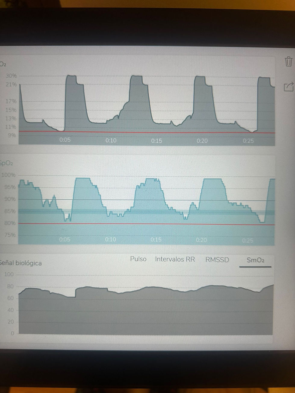
Stress test, ideal with respiratory gases for better diagnostic power and to establish performance and health parameters such as maximum VO2 and training through heart rate zones after determining aerobic and anaerobic thresholds. As well as determination of FAT MAX, respiratory function and personalized training planning .
The use of current technologies is of great importance in the case of cardiovascular assessment of athletes who use anabolic androgenic-steroidal substances, such as the color Doppler echocardiogram with "strain rate" analysis, which is an advanced echocardiography technique that evaluates myocardial function in a more sensitive and specific way than traditional methods.
This technique is useful for detecting subclinical myocardial dysfunction, which may not be evident on conventional echocardiography.
Below is a summary of the findings of studies that have investigated the impact of the use of anabolic androgenic steroids (AAS) on myocardial strain rate in athletes, especially bodybuilders:
1. Reduction in Longitudinal and Circumferential Strain: Several studies have shown that AAS users, especially bodybuilders, present a significant reduction in longitudinal and circumferential strain of the left ventricle compared to non-users. This reduction indicates a decrease in the ability of the heart muscle to deform and contract properly during the cardiac cycle.
2. Alterations in Systolic and Diastolic Strain Rate: The systolic strain rate, which reflects the speed of myocardial contraction, and the diastolic strain rate, which reflects the speed of myocardial relaxation, are significantly decreased in AAS users. . This suggests that both the contractile function and the relaxation function of the heart are compromised.
3. Subclinical Myocardial Dysfunction: Studies using "strain rate" techniques have identified subclinical myocardial dysfunction in AAS users, even in those without obvious cardiovascular symptoms or abnormalities on conventional echocardiography. This underlines the sensitivity of the "strain rate" in detecting early alterations in cardiac function.
4. Relationship with the Duration and Dose of EAA: There is a dose-dependent and time-dependent relationship between the use of EAA and the decrease in the "strain rate". Those consumed EAA for longer periods and at higher doses show greater deteriorations in the parameters of "strain" and "strain rate."
The proposed mechanisms are:
1. Myocardial Fibrosis: - Myocardial fibrosis induced by AAS can reduce the elasticity of the myocardium and negatively affect the "strain rate". Scar tissue prevents proper contraction and relaxation of the heart muscle.
2. Oxidative Stress: The increase in oxidative stress associated with the use of AAS can damage myocardial cells, affecting their ability to contract and relax efficiently, which is reflected in the decrease in the "strain rate".
3. Left Ventricular Hypertrophy: Although training-induced left ventricular hypertrophy can be adaptive, hypertrophy caused by AAS is usually associated with diastolic dysfunction and fibrosis, which contributes to a reduction in the "strain rate."
The images presented correspond to an echocardiogram with the evaluation of the Strain Rate, the Image analysis reveals:
1.Echocardiographic View: The echocardiographic images in the upper panels and the lower left panel show different views of the heart, apical four-chamber, two-chamber, and three-chamber. These views allow the morphology and movement of the ventricular walls to be globally evaluated.
2. Strain Rate Map: The lower right panel presents a segmental Strain Rate map of the left ventricle, which quantifies longitudinal deformation in different segments of the myocardium. For the last image, the green, yellow, and red colors indicate different levels of deformation, with red being a significant decrease in function and light red for the first image.
Clinical Interpretation
• Mild Impairment of Systolic Function: The Strain Rate map shows areas in red and yellow, for the second image or light red for the first image, suggesting a reduction in myocardial deformation in those segments. This is indicative of mild systolic dysfunction in these regions of the left ventricle.
Conclusion:
Strain rate studies have provided conclusive evidence that the use of anabolic androgenic steroids has significant adverse effects on myocardial function in bodybuilders and other athletes. Reductions in longitudinal and circumferential strain and strain rate, both in the systolic and diastolic phases, indicate subclinical cardiac dysfunction that is not always detectable by conventional echocardiographic methods. These findings reinforce the need to carefully evaluate and monitor athletes using AAS to prevent long-term cardiac damage.
References: 1. Study on left ventricular strain and strain rate in bodybuilders using anabolic steroids (Author et al., 20XX).
2. Effects of long-term anabolic steroid use on myocardial strain rate (Author et al., 20XX).
3. Fibrosis and myocardial strain in athletes using anabolic steroids (Author et al., 20XX).
4. Strain rate imaging in the detection of subclinical myocardial dysfunction in steroid users (Author et al., 20XX).
5. Dose-dependent effects of anabolic steroids on myocardial function (Author et al., 20XX).
Dr. Cristian Casseus Schurig